.JPG)
Exploring the world of microfluidics at VIB
The FabLab collaboration with KU Leuven allows VIB researchers to assess the potential of microfluidics for their projects.
The power of miniature channels
Imagine a world where a drop of liquid holds the key to unlocking mysteries at the cellular level or where complex biochemical reactions occur within a network of channels smaller than a human hair. This world is called microfluidics.
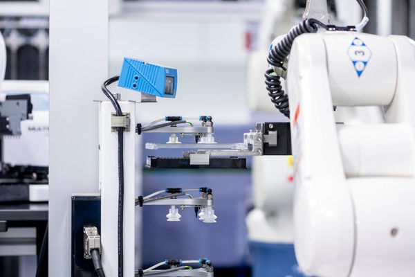
Microfluidics is a cutting-edge technology that enables scientists to manipulate fluids with unprecedented control and accuracy. Cleverly designed circuits play a pivotal role in microfluidics and their use ranges from healthcare diagnostics to environmental monitoring.
While the demand for microfluidic devices is soaring, the development and fabrication process require specialized expertise and infrastructure – there is no ‘one-size-fits-all’ solution. To meet this demand, VIB has been institutionally collaborating with the Biosensors lab of Jeroen Lammertyn at KU Leuven from 2020 onward. This partnership gives VIB teams access to the know-how needed to design their own microfluidic circuits. Projects are selected based on several criteria, including novelty, impact on VIB research, diversity of projects, complexity, and available capacity.
Iene Rutten, researcher in the Lammertyn lab who works on the VIB projects, on the partnership: "For us, this is a win-win collaboration. We can offer our technology and expertise to solve problems in the life sciences, but at the same time, we are challenged to develop new microfluidic chip designs and microfabrication protocols. With this initiative, we can accelerate the implementation of microfluidics as a key enabling technology to advance life science research."
Enabling science
One of the fields where the VIB collaboration with the Lammertyn lab is making a big impact is plant research. In the past, vertical imaging of roots was impossible. But now, through optimizing RootChip technology, the labs of Daniël Vandamme and Moritz Nowack achieve a unique view of developing roots, which allows them to test the effects of specific compounds and environmental conditions on the health and growth of seedlings.
Evelien Mylle from the Van Damme lab: “Our microfluidic setup at PSB allows us to monitor the immediate effect of compound treatments on root dynamics (growth, marker expression) in real-time. Plant roots are grown in a RootChip, through which the fluid flow is controlled by a microfluidic system that is mounted on a confocal microscope. This setup allows the roots to grow along the gravity vector and we can follow the roots over an extended period of time (days) using a root tip tracking system.”
.JPG)
.jpeg)
Left: the microfluidic setup at PSB; right: detail of the RootChip.
Another research area in which VIB research meets microfluidics is microbiology. The lab of Jeroen Raes at the VIB-KU Leuven Center for Microbiology has long been working on isolating and characterizing the diversity of different human microbial ecosystems, with the gut microbiome being the most famous example. However, current bulk approaches tend to filter out the rare and slow-growing microbiome members. To unlock that microbial dark matter, the team is working on the optimization of microfluidics-based workflows for the targeted and untargeted isolation of anaerobic bacteria from human fecal samples.
Geert Huys, from the Raes lab: “Despite their revolutionary contribution to the human microbiome field, most ‘omics’ technologies can't discriminate and validate the function of individual microorganisms. To try and capture this 'microdiversity' at maximal resolution, microfluidics-based single-cell analyses hold the potential to address novel biological questions at the finest functional level of gut ecosystems using cellular, metabolic, DNA, and RNA readouts.”
Research into human health and disease benefits from microfluidics too. The Switch lab at the VIB-KU Leuven Center for Brain & Disease Research, led by Frederic Rousseau and Joost Schymkowitz, uses microfluidics to improve the diagnosis of diseases marked by issues with protein aggregation, such as major neurodegenerative diseases like Alzheimer’s and Parkinson’s disease.
Nikolaos Louros from the Switch lab explains: “The cornerstone of this project is developing a microfluidics-based digitized aggregation assay (DIGAS) to quantify particles that can ‘seed’ protein aggregation, derived from challenging samples (patient tissue extracts, cerebrospinal fluid, blood). Specific dyes that fluoresce in the presence of formed aggregates then allow us to measure the seeding reactivity with unprecedented accuracy and sensitivity. Due to this extreme sensitivity, this method can lead to a future diagnostic platform for different brain disorders and disease progression stages.”

The lab of Jan Cools at the VIB-KU Leuven Center for Cancer Biology is also looking into the potential of microfluidics for their work. More specifically, they want to develop a microfluidics device to detect the characteristics of white blood cells in blood samples.
Krista Verhoeft from the Cools lab: “A first application will be to detect abnormalities in blood samples in a rapid and simple manner. The second application will be to predict the drug sensitivity of leukemia cells in patients diagnosed with hematological malignancy. Usually, these tests require time, several medical professionals, and extensive laboratory analyses. Our microfluidics devices would significantly reduce the time and personnel required and be both user-friendly and fast. In the future, this might enable rapid detection of abnormalities in blood samples at a doctor's appointment, and potentially in developing countries where access to medical health care professionals is limited.”
Enabling technology
Microfluidics is not only a technology that enables science; it’s also a technology that enables further technology development. Several VIB Core Facilities, for example, are exploring microfluidics to expand their service offering. The VIB Bioimaging Core Leuven is looking into microfluidics to develop multimodal imaging of the same sample at different scales, which would overcome a crucial problem of relating the micro- to the mesoscale: connect our understanding at the micro- and mesoscale and so overcome the central challenge in imaging: the problem of resolution versus sample size.
Sebastian Munck, innovation technologist at the Bioimaging Core, explains: “Our goal is to build a phantom, which is a 3D test sample used in medical imaging to simulate the imaging properties of (human) tissue and quality control the imaging devices. Combining a 3D-printed phantom with microfluidics will effectively give us a multimodal phantom that allows us to test and validate various imaging approaches that will ultimately be a key step in generating multimodal datasets in the various research areas studied at VIB.”

.png)
Left: the advantages of multimodal imaging; right: droplet imaging with DE-FACS.
Gert Van Isterdael, head of the VIB Flow Core, agrees with Sebastian on the huge potential of microfluidics as an enabling technology.
“We are looking into how to use microfluidics to optimize double emulsion fluorescence-activated cell sorting or DE-FACS. In short, we are using both conventional and image-enabled FACS sorters to separate droplets containing cells or molecules of interest at high speed. We believe that combining microfluidics with DE-FACS can make this process faster and more accurate, greatly benefiting many VIB research groups.”
From protein aggregates to roots, and from blood cells to microbes, microfluidics has certainly proven its use at VIB and its VIB Technologies’ Core facilities thanks to the collaboration with the KU Leuven team of Jeroen Lammertyn.
We can’t wait to see what the future brings!
If you have any questions on the use of microfluidics at VIB or see potential to use it in your own research, feel free to contact VIB Technologies.
Want to be kept up-to-date on our biotechnological news and stories? Join our community and subscribe to our bi-monthly newsletter.
Gunnar De Winter