From live action to the molecular backstory of single cells
Tech Watch scouts the wave of new platforms targeting live single-cell characterization
Single-cell biology has long forced researchers into an uncomfortable trade-off: watch what a cell does, or find out what it expresses, but rarely both on the same cell. Most tools capture static snapshots of inherently dynamic processes, leaving a gap between observed behavior and molecular understanding. The VIB Tech Watch Core, always on the hunt for technologies that can push VIB research forward, has tracked the sprouting platforms aiming to bridge that gap.
Biology is dynamic. Cells move, change shape, secrete signals, kill neighbors, and respond to their environment. But most of our tools for studying this behavior force us to work with static snapshots. We fix cells to study morphology. We collect supernatants at a single time point to measure what they have secreted. We lyse cells to sequence nucleotides. We take one image and treat it as the definitive shape of a cell that, in reality, has been shifting and migrating the whole time.
Rather than focusing solely on molecular snapshots obtained from such destructive endpoint assays, the latest live single-cell characterization platforms aim to preserve cells in defined micro-environments and monitor their behavior over time. In some cases, they even enable the retrieval of individual cells for downstream analyses. For many VIB research groups, this shift opens new experimental possibilities at the interface of cell biology, immunology, functional genomics, and translational research.
The shared workflow behind most platforms
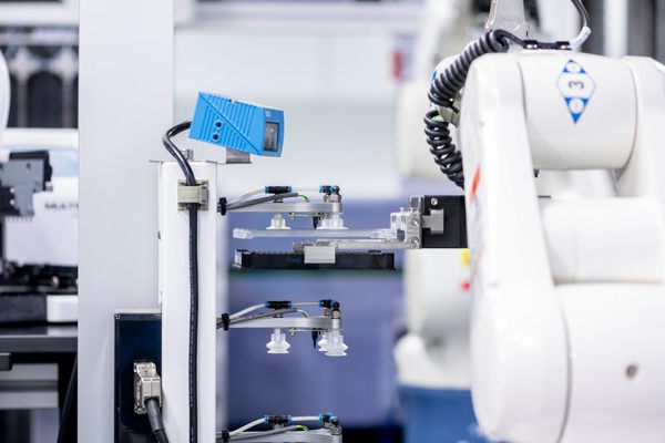
At the core of these platforms lies a shared conceptual workflow. Individual cells are first compartmentalized. Within these confined spaces, cells can be cultured alone or in controlled cocultures, exposed to perturbations such as drugs, antibodies, or target cells, and followed by live imaging. Integrated readouts commonly include brightfield and fluorescence microscopy, often combined with automated image analysis to quantify dynamic features such as cell morphology, migration, secretion, or cytotoxic activity.
A key ambition of many systems is to connect these functional phenotypes back to molecular information, either by retrieving selected cells alive for further processing or by linking imaging data to sequencing-based readouts.
The key differentiator between platforms is how they compartmentalize cells, and what that implies for throughput, assay flexibility, and cell retrieval.
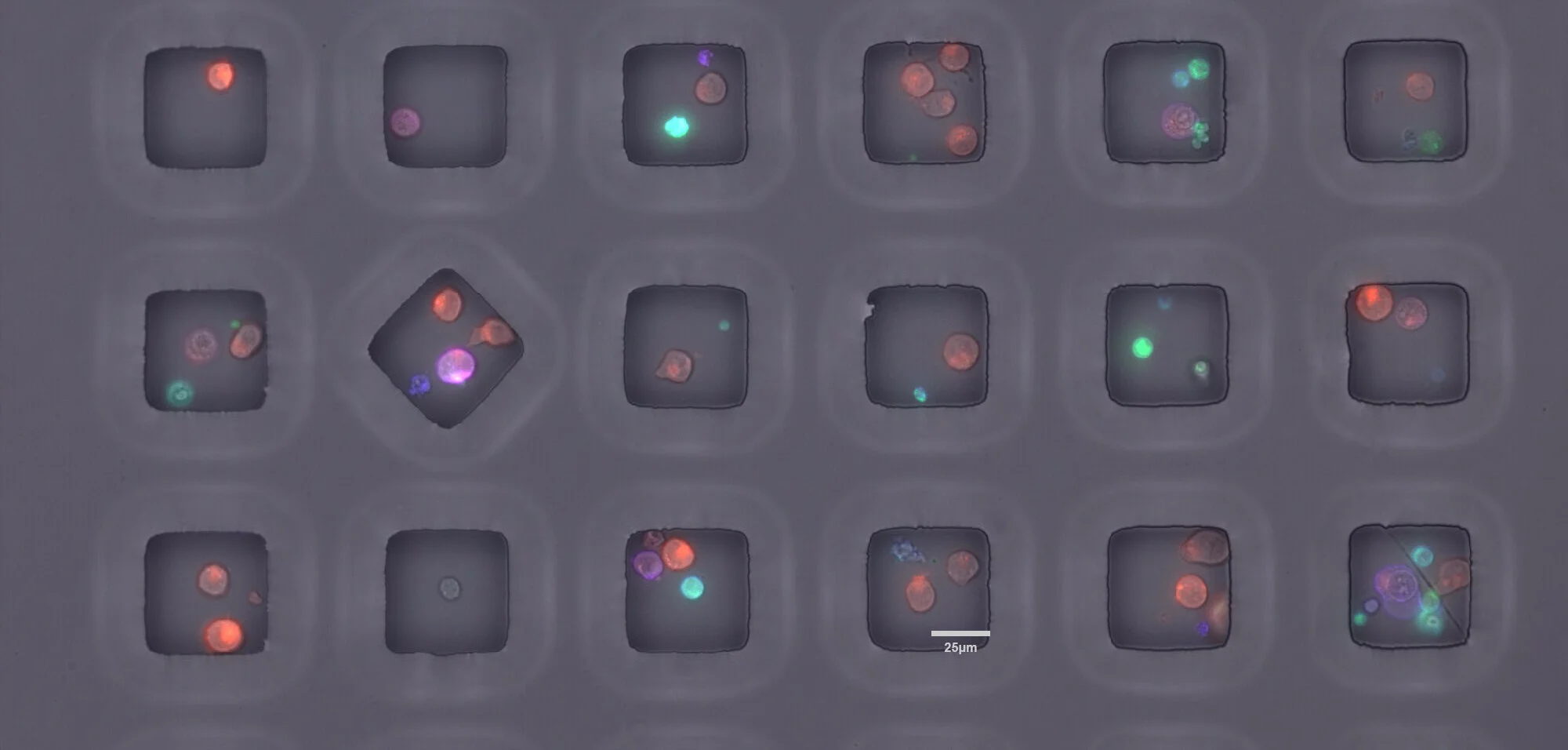
Microwells, nanowells, and picowells as addressable ‘mini labs’ for single cells
A first approach relies on dense arrays of microwells or nanowells fabricated in glass or polymer substrates. Cells are dispensed into these wells, often at nanoliter volumes, and imaged over time under controlled environmental conditions.
Technology perks ✨
Because each well has a fixed spatial address, individual cells or small cell groups can be tracked across hours or days. Some systems also include dedicated extraction mechanisms that allow the contents of selected wells to be recovered while maintaining cell viability.
Platforms on Tech Watch’s radar
Celldom offers a microwell-based live single-cell assay platform, CloneXplorer, that combines long-term time-lapse imaging with AI-driven analysis and on-instrument retrieval of selected cells for downstream expansion or sequencing. Tech Watch is in close contact with the Celldom team and is tracking the platform’s development and fit for VIB use cases.
CellChorus focuses on dynamic profiling of cell behavior using Timelapse Imaging Microscopy in Nanowell Grids (TIMING), emphasizing quantification of migration, contact dynamics, cytotoxicity, and survival, with the possibility to link functional performance to downstream molecular analyses.
Tech Watch has evaluated the CellShepherd® platform from Arralyze in 2024. This integrated workstation combined single-cell dispensing into glass nanowell arrays with live imaging and a cell extraction unit, enabling functional screening of cells of interest.
Other picowell-based systems focus on capturing rich imaging data and associating it with molecular information through spatial or optical barcoding strategies.
In the FLEX picowell chip from Flexomics, tens to hundreds of thousands of cells are captured in microscope-compatible arrays containing preloaded, barcoded beads. Cells can be imaged over time and subjected to functional assays, after which they are lysed in situ and their transcripts captured on the beads. Sequencing data can then be computationally linked back to imaging-derived phenotypes. Tech Watch is currently in contact with Flexomics and looking into evaluating their technology together with dedicated VIB researchers.

Droplet microfluidics for programmable microreactors at scale
A second strategy is to encapsulate cells in picoliter droplets that are actively filtered to enrich for single-cell occupancy before being immobilized on addressable surfaces. Using light-controlled electrowetting (oEWOD), droplets can be moved, merged, incubated, and imaged in a programmable manner.
Technology perks ✨
This approach enables complex, multistep functional assays to be performed sequentially on thousands of individual cells in parallel. Selected droplets can subsequently be dispensed into standard well plates, preserving the link between functional readout and physical samples for follow-up analyses such as cell culture or sequencing.
On Tech Watch’s radar
Envisia, developed by Lightcast, is currently available in the Tech Watch demo lab in Leuven. This technology is of particular interest for studying cell-cell interactions and functional heterogeneity in a highly controlled droplet environment, while allowing retrieval of cells of interest in microtiter plates.

Optofluidic precision manipulation with exportable cells
A third approach uses optofluidics to position and culture cells in small compartments (Nanopens) for longitudinal observation. Reagents can be delivered in highly controlled ways, and multiple assays can be performed sequentially on the same cells.
Technology perks ✨
A defining feature is the ability to export specific selected cells after on-chip characterization, directly connecting performance to downstream genomic/transcriptomic analysis. While you can also do this with droplet microfluidic platforms (above), the fixed nanopen compartment offers more flexibility for downstream cell selection.
The Beacon Discovery system from Bruker offers varying levels of throughput and flexibility.

On-demand “cages” keep cells fixed and biology intact
An alternative approach is to first image cells in suspension and then enclose them on demand within microfabricated, three-dimensional structures.
Technology perks ✨
This strategy is particularly attractive for studying adherent cells or complex cell-cell interactions over extended periods, as it preserves lineage continuity.
Tech Watch has been piloting the Cellanome R3200 in the Tech Watch Ghent demo lab. This platform uses light-initiated polymerization to form so-called CellCage enclosures around selected cells, including adherent and large cells or complex mixtures. These cages are permeable to nutrients and reagents while keeping cells spatially fixed, enabling longitudinal studies without repeatedly moving samples between instruments. The system integrates automated imaging, segmentation, and cage fabrication into a single workflow. Following imaging, it supports barcoded capture of RNA and/or CRISPR tags to link molecular profiles directly to imaging-derived phenotypes.

Flow-cell-scale multimodal profiling
At the other end of the spectrum are platforms designed for very large-scale profiling of cells directly on flow-cell-like substrates. These systems blur the line between imaging and sequencing by performing both operations on the same surface.
AVITI24™ from Element Biosciences (available through the VIB Nucleomics Core), which combines cellular imaging with sequencing-based detection of RNA and protein targets in situ. Such approaches enable multimodal profiling of very large numbers of cells in a single run.
Minos Biosciences is developing a microfluidic platform that couples high-throughput live-cell imaging of complex cell-pathogen or cell-cell interactions with sequencing-based molecular profiling, aiming to resolve dynamic functional states and their molecular drivers in parallel.
Zafrens’ Z-ScreenTM technology condenses 50-200K indexed micro‑experiments onto a single array. It pairs time‑lapse imaging with single‑cell multi‑omics and supports large perturbation libraries, enabling simultaneous functional and molecular readouts from the same cells.

Across all these platforms, Tech Watch consistently encounters similar tradeoffs. Systems that offer fine-grained control over live cells or enable recovery for downstream assays often come with increased instrumental complexity and lower raw throughput. Platforms optimized for scale and multimodal profiling may sacrifice the ability to retrieve living cells. Single-cell retrieval, particularly when combined with robust and standardized sequencing workflows, remains one of the more challenging aspects.
Tech Watch also monitors several other technologies that approach live single-cell analysis from complementary angles.
Cellply’s VivaCyte platform uses dense microfluidic coculture arrays and automated imaging to assess immune cell potency and phenotype at single-cell resolution, primarily in the context of cell therapy characterization, with a focus on standardized, end-to-end workflows rather than live cell recovery.
DPBio offers droplet-based microfluidic systems for ultra-high-throughput single-cell screening, commonly applied to antibody discovery or enzyme screening, where functional readouts are captured within droplets and hits are selected based on activity.
One Biosciences applies single-cell transcriptomic profiling combined with AI-based analysis to resolve tumor heterogeneity from clinical samples, prioritizing large-scale molecular characterization over live cell manipulation or retrieval.
Together, these technologies further illustrate the breadth of approaches currently emerging in the live single-cell analysis space, spanning dynamic functional imaging, high-throughput screening, and large-scale molecular profiling, each with distinct trade-offs in terms of resolution, scalability, and downstream compatibility.
This overview is not exhaustive: the landscape is moving fast, and new platforms appear continuously. If you know of a promising technology or company we should have on our radar (or include next time), feel free to reach out.
If you have a research question where linking dynamic phenotype to molecular readouts could be decisive, reach out to Tech Watch to discuss fit and potential access routes.


.jpeg)

.jpeg)

VIB Tech Watch | Disclaimer
The role of Tech Watch is not to endorse specific technologies, but to help VIB researchers navigate this rapidly evolving landscape. By scouting emerging platforms, evaluating them based on concrete research needs, and organizing hands-on pilots where possible, the team aims to connect research projects to the most compatible platform and lower the barrier for adopting novel experimental approaches. Extensive evaluation reports are available for the VIB researchers to make an informed decision on which platform to pursue.